43 microscope images with labels
Amazon.com: My First Lab Duo Scope Microscope - Young … KIDS MICROSCOPE KIT -- This kids microscope kit, in addition to the user manual and experiment guide, includes 5 blank slides, 1 concavity (well) slide, 4 prepared slides, 2 bottles of stain, 5 slide labels, 5 cover glasses, 50 sheets of lens paper, 1 plastic transfer pipette, 1 plain wooden applicator, 1 cotton-tipped applicator, 1 plastic forceps, 1 plastic test tube and 1 plastic … 26+ Picture Of A Microscope With Label PNG 26+ Picture Of A Microscope With Label PNG. Microscopes are specially created to magnify the image of the subject being studied. Students label the parts of the microscope in this photo of a basic laboratory light microscope. Microscope Drawing And Label at GetDrawings | Free download from getdrawings.com I searched for this on bing.com/images.
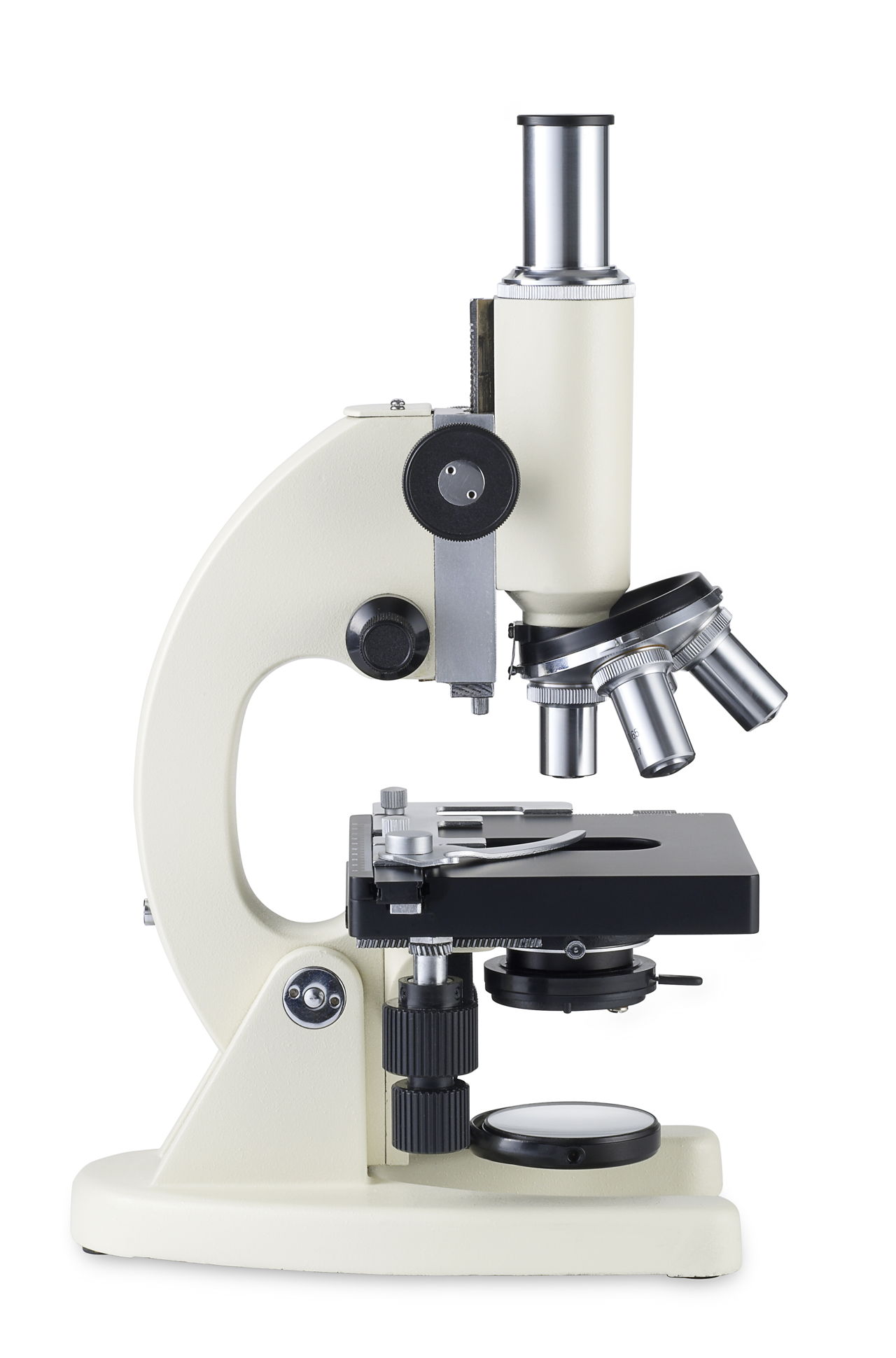
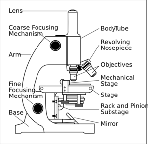
16 Parts of a Compound Microscope: Diagrams and Video It's actually not a toy microscope, it's a functional microscope that produces great images for the price. I bought it for less than $100 dollars but you can check the current price on Amazon. 1. Head (Body) The head, also referred to as the body of the microscope, is a structural component that contains the optical parts of the microscope.

Microscope images with labels
Microscope Drawing And Label - Painting Valley Are you looking for the best images of Microscope Drawing And Label? Here you are! We collected 33+ Microscope Drawing And Label paintings in our online museum of paintings - PaintingValley.com. ADVERTISEMENT LIMITED OFFER: Get 10 free Shutterstock images - PICK10FREE label microscope diagram compound parts light labeling functions microscopic Microscope Slide Label Templates - TemplateMonster Minimalist is a beautiful PowerPoint template you can use to design professional presentations for business and agency meetings and events. The template comes with 140 unique slides in HD resolution and features image placeholders, world maps, icons, and much more.Key Features : 140 different slides, Simple. Explanation and Labelled Images - New York Microscope Company The samples are labeled with fluorophore where they absorb the high-intensity light from the source and emit a lower energy light of longer wavelength. The resulting fluorescent light is then separated from the surrounding radiation with filters, allowing the observer to see only the fluorescing material.
Microscope images with labels. Parts of Stereo Microscope (Dissecting microscope) - labeled diagram ... Stereo microscopes (also called Dissecting microscope) are branched out from other light microscopes for the application of viewing "3D" objects. These include substantial specimens, such as insects, feathers, leaves, rocks, sand grains, gems, coins, and stamps, etc. Functionally, a stereo microscope is like a powerful magnifying glass. Microscope labels | Bill Todt Microscope labels. Unlabelled image: Lab Index: 101 Class Page: Identify the following parts of the microscope: Eyepieces Observation tube Nosepiece Objectives Stand Specimen holder Mechanical stage Stage controls Condenser Iris diaphragm Coarse focus Fine focus Microscope With Labels clip art | Microscope parts, Scientific method ... Jul 3, 2012 - Download Clker's Microscope With Labels clip art and related images now. Multiple sizes and related images are all free on Clker.com. Parts of a microscope with functions and labeled diagram Optical parts of a microscope and their functions The optical parts of the microscope are used to view, magnify, and produce an image from a specimen placed on a slide. These parts include: Eyepiece - also known as the ocular. This is the part used to look through the microscope. Its found at the top of the microscope.
Smart Microscope for Lab Routine and Research - ZEISS In the past, documenting samples with multiple fluorescent labels in your routine lab could be time consuming. To get best image quality, you needed to manually switch filters, adjust illumination intensities and exposure times and to snap each single channel image. For four different channels, this could sum up to 15 steps and clicks. With Smart Microscopy, this is a … Parts of Stereo Microscope (Dissecting microscope) – labeled … Compared to a compound microscope where the objectives attached to the nosepiece can be seen and identified individually (based on color bands and their respective labels), the objectives of a dissecting microscope are located in a cylindrical cone and, therefore, are not directly seen. For the stereo microscope that comes with multiple objective lens sets (fixed power style), the … Parts of the Microscope with Labeling (also Free Printouts) Microscopes are specially created to magnify the image of the subject being studied. This exercise is created to be used in homes and schools. the microscope layout, including the blank and answered versions are available as pdf downloads. Click to Download : Label the Parts of the Microscope (A4) PDF print version. Skin Images Labeled | Virtual Anatomy Lab VAL - ncccval Skin Images Labeled | Virtual Anatomy Lab VAL ... Connective Tissue Images Unlabeled. Microscope. Microscope Images Labeled. Microscope Images Unlabeled. Mitosis. Mitosis Images Labeled. Mitosis Images Unlabeled. Skin. Skin Images Labeled. Skin Images Unlabeled. Skeletal system. Skeletal Images Labeled. Skeletal Images Unlabeled.
Microscope Parts, Function, & Labeled Diagram - slidingmotion Microscope parts labeled diagram gives us all the information about its parts and their position in the microscope. Microscope Parts Labeled Diagram The principle of the Microscope gives you an exact reason to use it. It works on the 3 principles. Magnification Resolving Power Numerical Aperture. Parts of Microscope Head Base Arm Eyepiece Lens Microscope With Labeled Parts And Functions The optical parts of the microscope are used to view, enlarge, and produce an image from a sample placed on a slide. These parts include. Eyepiece: Eyepiece also contains ocular lens. It enhance the image of the viewer. This part is used for checking through the microscope. Eyepiece is found at the upper part of it. Microscope Labeled Pictures, Images and Stock Photos Browse 48 microscope labeled stock photos and images available, or start a new search to explore more stock photos and images. Newest results Fluorescent Imaging immunofluorescence of cancer cells growing... Plant Tissue Systems vector illustration. Labeled biology... Microscope diagram vector illustration. Labeled zoom instrument... Microscope Labeling - The Biology Corner The google slides shown below have the same microscope image with the labels for students to copy. I often spend the first day walking students through the steps and having them look at a single slide as we do the steps. Students are often very enthusiastic about using microscopes and will try to start with the high power objective.
Biology Eleven: Looking at Agiospermae Reproductive organs. Also examine the leaf and stem cross ...
Canon U.S.A., Inc. | EOS Utility EOS Utility is an application that brings together functions to communicate with the camera. These functions include downloading and displaying images, remote shooting, and camera control for each setting. For download instructions follow the steps below. Have your camera's Serial Number ready before you begin.

Broad mite feeding injury on New Guinea impatiens | UMass Center for Agriculture, Food and the ...
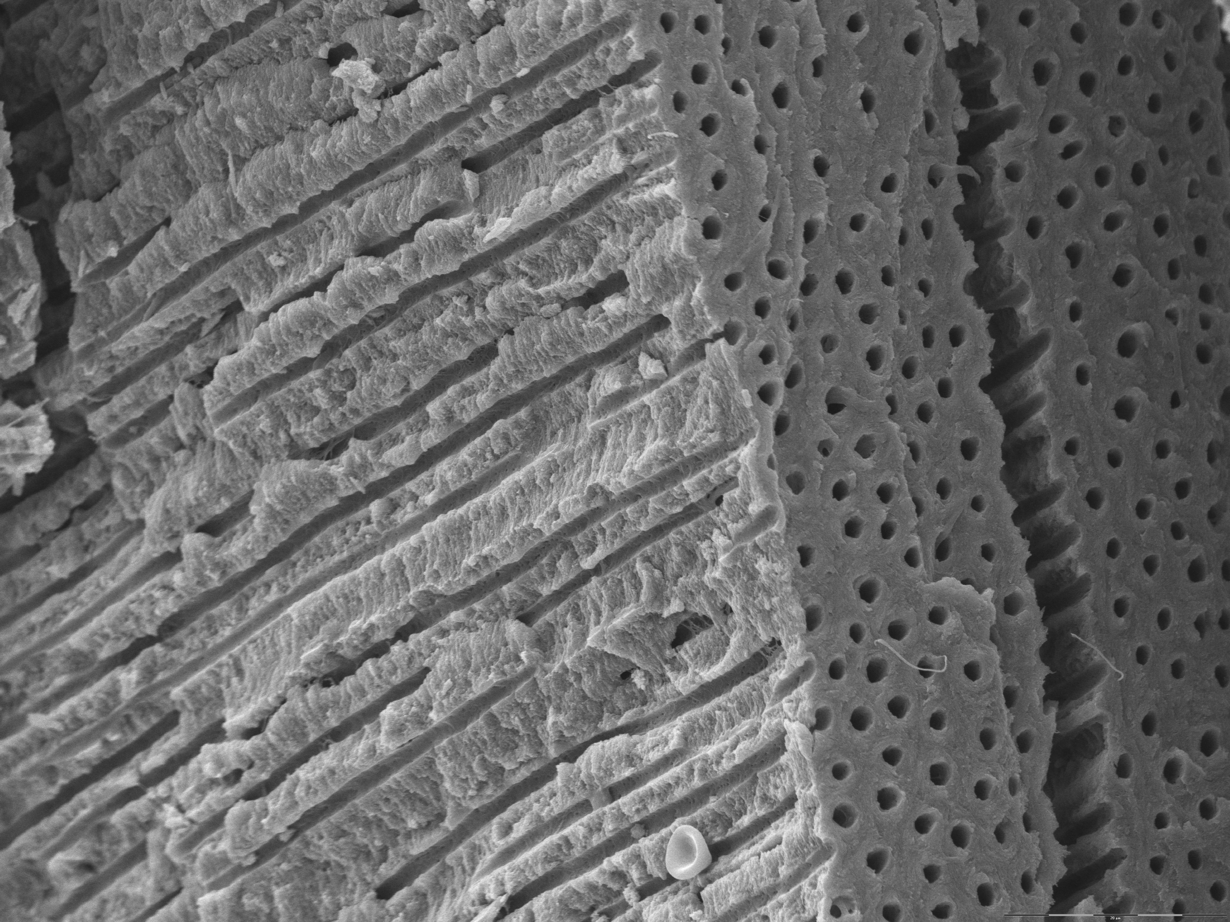
Amazing 27 Things Under The Microscope With Diagrams Skeletal muscle under the microscope 40X magnification 100X magnification 400X magnification 20. Skin under the microscope 21. Snowflake under the microscope 22. Sperm under the microscope Direct observation Observation after staining 23. Spirogyra under the microscope 24. Virus under the microscope Fluorescence microscope
Dentine with dentinal tubules - Anatomicum.com
Adipose tissue microscope Images, Stock Photos & Vectors | Shutterstock Find Adipose tissue microscope stock images in HD and millions of other royalty-free stock photos, illustrations and vectors in the Shutterstock collection. Thousands of new, high-quality pictures added every day. Images. Footage. Music. Templates. Editorial. 3D Models. Tools. Blog. Enterprise. Pricing. Menu. Image Adipose tissue microscope ...

Label the Microscope Part
Histology - Yale University Answer: The kidneys would appear the same under the light microscope, but the foot processes of the podocytes would be missing in the EM of the minimal change kidney. Patients with this disease have edema because they can no longer repel proteins from entering the urine, and there is a loss of albumin from the blood into the urine, which is excreted. Albumin normally …
Label a microscope - Teaching resources
Label the microscope — Science Learning Hub All microscopes share features in common. In this interactive, you can label the different parts of a microscope. Use this with the Microscope parts activity to help students identify and label the main parts of a microscope and then describe their functions. Drag and drop the text labels onto the microscope diagram.
33 Label A Compound Light Microscope - Labels Database 2020
300+ Free Microscope & Laboratory Images - Pixabay Upload 399 Free images of Microscope Related Images: laboratory science bacteria research scientist lab biology chemistry medical Find your perfect microscope image. Free pictures to download and use in your next project. 399 Free images of Microscope / 4‹ ›

Give the label each part of the microscope: - Brainly.ph
Microscope Types (with labeled diagrams) and Functions This is an advanced microscope that has specific application in viewing, observing and measuring the optical thickness and phase of completely transparent specimens and objects. A tiny interferometer is used and a specimen is placed on beam path of it. This path is split and then rejoined to create two superimposed images of the specimen in focus.

Microscope labeling
Microscope picture label Flashcards | Quizlet Microscope picture label Flashcards | Quizlet Microscope picture label STUDY Flashcards Learn Write Spell Test PLAY Match Gravity Created by kfire Terms in this set (12) Arm What is the part labelled C? Base What is the part labelled D? Body tube What is the part labelled B? Ocular lens What is the part labelled A? Illuminator
34 Label Of A Microscope - Labels For You
Electron Microscopy Images - Dartmouth We have a library of images recorded using our scanning and transmission electron microscopes. Some are shown below and others elsewhere. These images are in the public domain. If you have questions about the images or want some specific images contact Max Guinel . Hibiscus Flower (August 2021) Morphy Amorphophallus titanum anther cross section.
2.3.3 Identify structures from electron micrographs of liver cells - YouTube
Compound Microscope Parts - Labeled Diagram and their Functions - Rs ... The eyepiece (or ocular lens) is the lens part at the top of a microscope that the viewer looks through. The standard eyepiece has a magnification of 10x. You may exchange with an optional eyepiece ranging from 5x - 30x. [In this figure] The structure inside an eyepiece. The current design of the eyepiece is no longer a single convex lens.

Synapse Science Magazine: Weird and Wonderful: Hidden Horrors Returns!
Microscope, Microscope Parts, Labeled Diagram, and Functions Microscope, Microscope Parts, Labeled Diagram, and Functions What is Microscope? A microscope is a laboratory instrument used to examine objects that are too small to be seen by the naked eye. It is derived from Ancient Greek words and composed of mikrós, "small" and skopeîn,"to look" or "see".
Microscope With Labels Clip Art at Clker.com - vector clip art online, royalty free & public domain
Labeling the Parts of the Microscope Labeling the Parts of the Microscope This activity has been designed for use in homes and schools. Each microscope layout (both blank and the version with answers) are available as PDF downloads. You can view a more in-depth review of each part of the microscope here. Download the Label the Parts of the Microscope PDF printable version here.
Organisation of cells animal tissues - Karnataka Open Educational Resources
Microscope Labeling - The Biology Corner Students label the parts of the microscope in this photo of a basic laboratory light microscope. Can be used for practice or as a quiz. ... 20. A microscope has an ocular objective of 10x and a high power objective of 50x, what is the microscope's total magnification? _____

Week 1: Microscope Usage & Snowflake Preservation - Inspiring a Love of Life-Long Learning
PDF Label parts of the Microscope Label parts of the Microscope: . Created Date: 20150715115425Z

Salisbury's Graduate Histology: Connective Tissue
Compound Microscope - Diagram (Parts labelled), Principle and Uses Compound Microscope - Diagram (Parts labelled), Principle and Uses As the name suggests, a compound microscope uses a combination of lenses coupled with an artificial light source to magnify an object at various zoom levels to study the object. A compound microscope: Is used to view samples that are not visible to the naked eye

Science labs - Akers 6th Grade Team
ZEISS Elyra 7 with Lattice SIM² Super-Resolution Microscope Images of Cos-7 cell stained with anti-alpha-Tubulin Alexa fluor 488 were processed with the conventional SIM algorithms based on generalized Wiener filter and with the novel SIM² reconstruction. The images show an improvement of resolution for SIM² compared to SIM. Objective: Plan-Apochromat 63× / 1.4 Oil

35 Label The Microscope Game - Labels For Your Ideas
Compound Microscope with labels Stock Vector | Adobe Stock Download Compound Microscope with labels Stock Vector and explore similar vectors at Adobe Stock. Adobe Stock. Photos Illustrations Vectors Videos Audio Templates Free Premium Editorial Fonts. ... Get 10 free Adobe Stock images. Start now. Get 10 free images. Unlock 200M+ assets in our full collection.

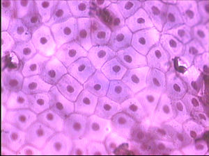
Microscopic Images
MINFLUX | Abberior Instruments Dyes & Labels; FAQ; Shop; MINFLUX . MINFLUX 3D. The MINFLUX platform offers an unprecedented array of imaging possibilities and allows you to resolve structures as small as a molecule, along all three dimensions. This unmatched resolution capability combined with unprecedented speeds reveals sample details never seen before. The MINFLUX is the world’s …
Post a Comment for "43 microscope images with labels"