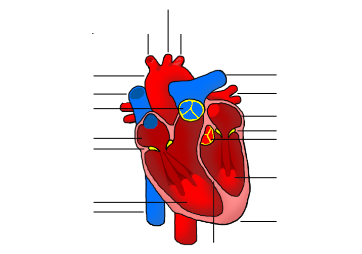
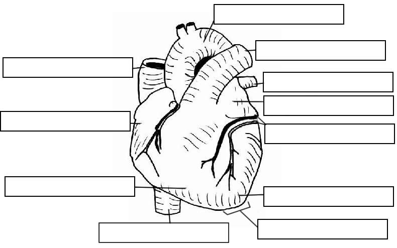
42 structure of the heart without labels
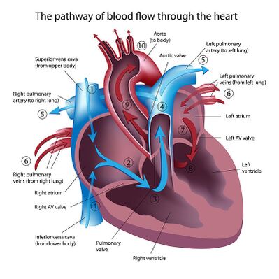
How the Heart Works: Diagram, Anatomy, Blood Flow - MedicineNet The heart is an amazing organ. It starts beating about 22 days after conception and continuously pumps oxygenated red blood cells and nutrient-rich blood and other compounds like platelets throughout your body to sustain the life of your organs.; Its pumping power also pushes blood through organs like the lungs to remove waste products like CO2.; This fist-sized powerhouse beats (expands and ... Cross Section of the Heart Diagram & Function | Body Maps - Healthline Cross-section. The chambers of the heart operate as a 'double-pump' system for the body's circulation. In coordination with valves, the chambers work to keep blood flowing in the proper ...
en.wikipedia.org › wiki › DapagliflozinDapagliflozin - Wikipedia Medical uses. Dapagliflozin is used along with diet, exercise and usually with other glucose lowering medications, to improve glycaemic control in adults with type 2 diabetes and to reduce the risk of hospitalisation for heart failure among adults with type 2 diabetes and known cardiovascular disease or other cardiovascular risk factors (including high blood pressure, high cholesterol and ...
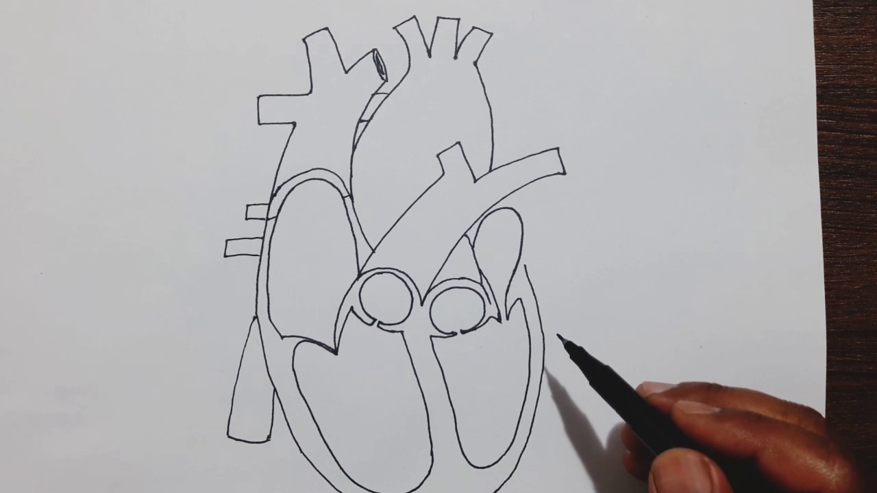
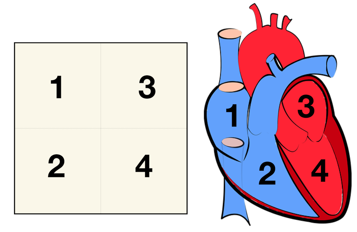
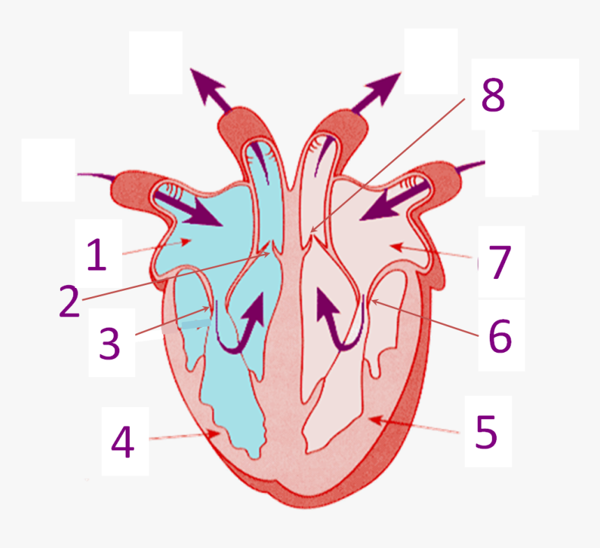
Structure of the heart without labels
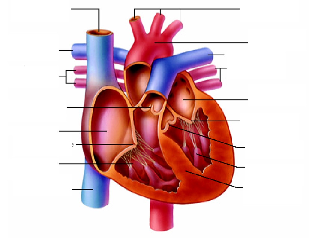
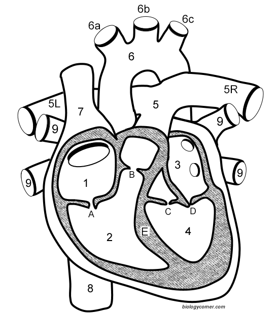
Structure and Function of the Heart - News-Medical.net Structure of the heart, The heart wall is composed of three layers, including the outer epicardium (thin layer), middle myocardium (thick layer), and innermost endocardium (thin layer). The... Diagram Of Fish With Label / External Morphology Of Rohu Fish With ... Plain diagram of the heart with labels to add and a cloze exercise on the pathway of blood through the heart. Structure of a typical fish (with diagram). A quality educational site offering 5000+ free printable theme units, word puzzles, writing forms, book report forms,math, ideas, lessons and much more. Anatomy of a Human Heart - U of M Health Located between the lungs in the middle of the chest, the heart pumps blood through the network of arteries and veins known as the cardiovascular system. It pushes blood to the body's organs, tissues and cells. Blood delivers oxygen and nutrients to every cell and removes the carbon dioxide and other waste products made by those cells.
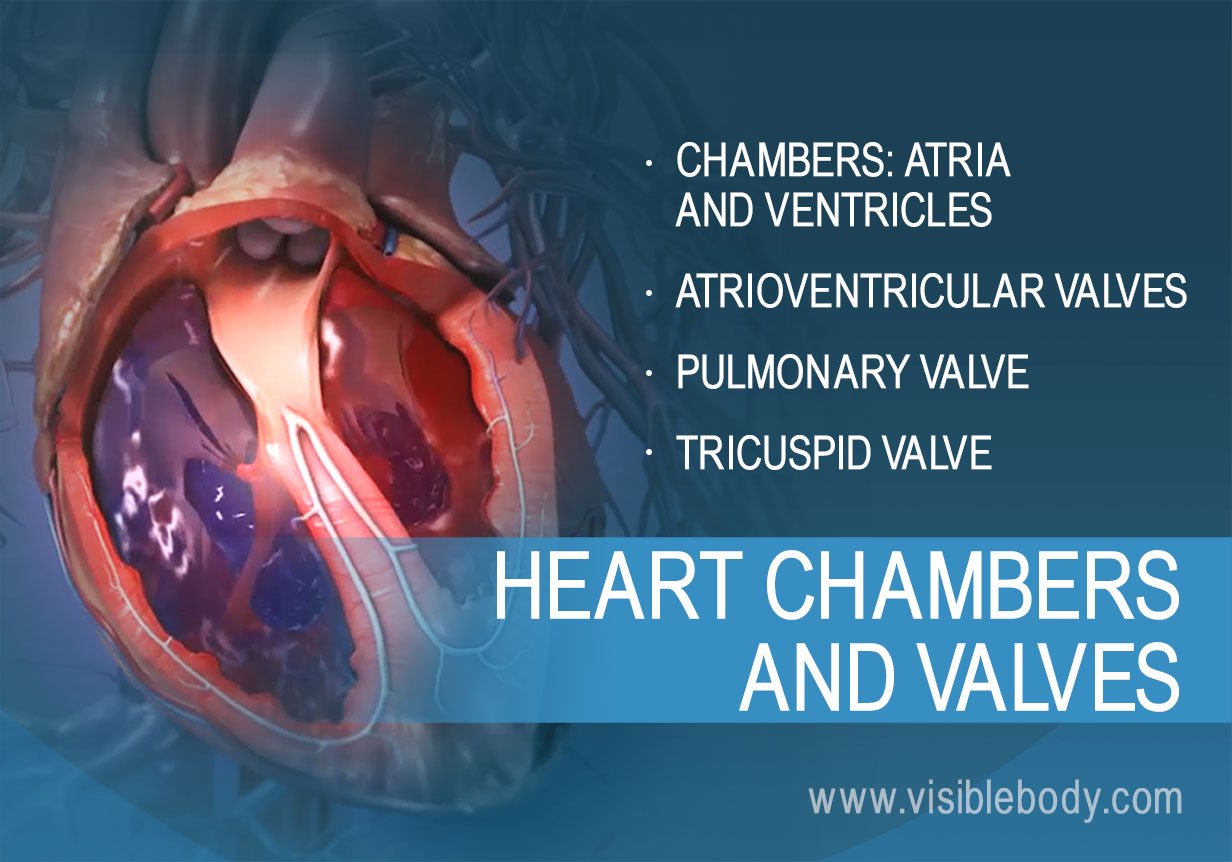
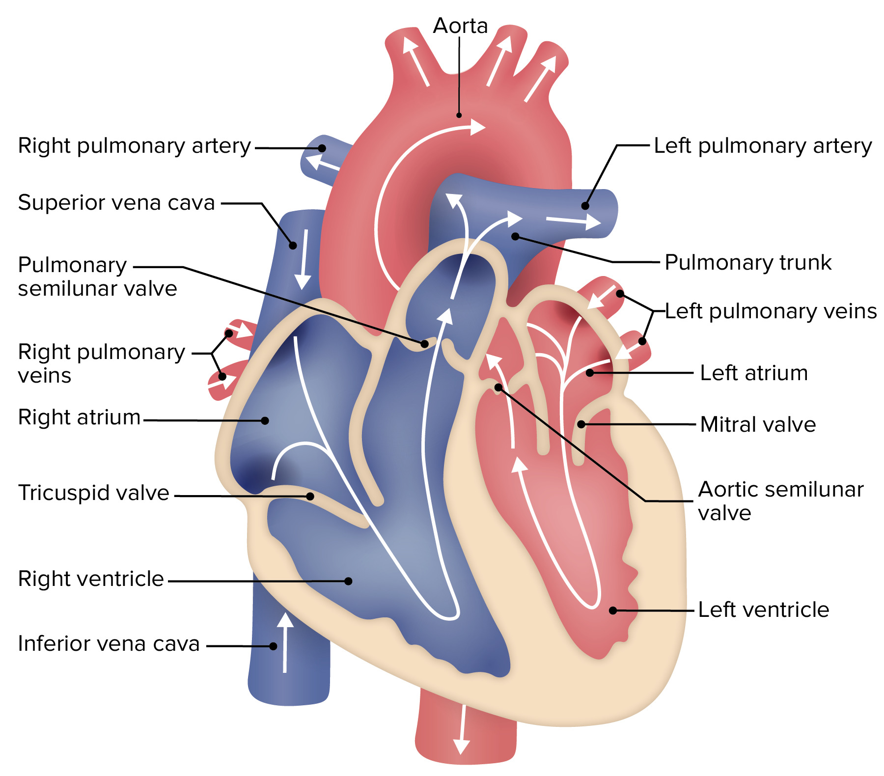
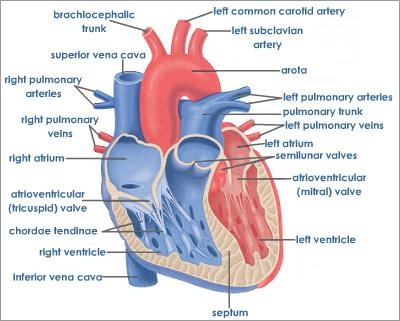
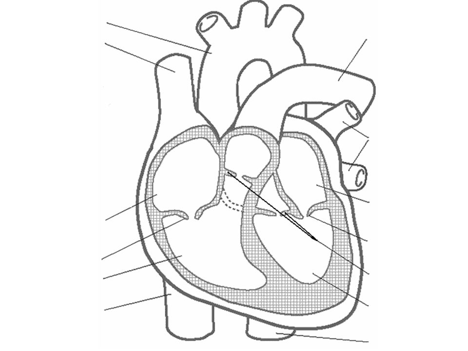
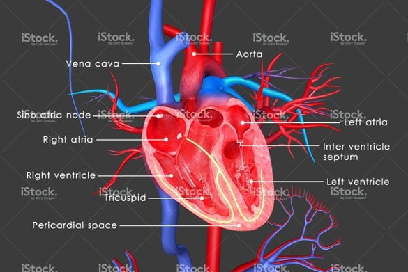
Structure of the heart without labels. Heart anatomy: Structure, valves, coronary vessels | Kenhub The heart is shaped as a quadrangular pyramid, and orientated as if the pyramid has fallen onto one of its sides so that its base faces the posterior thoracic wall, and its apex is pointed toward the anterior thoracic wall. heart diagram without labels circulatory blood system worksheet unlabeled vessels coloring diagram drawing anatomy circulation animals physiology heart flow body vessel colour printable wikieducator. The Heart rjoseph1994.blogspot.com. chambers. 30 Heart Diagram No Labels - Wiring Diagram Database kovodym.blogspot.com. heart diagram labels glogster edu drum human › en › healthy-livingCarbohydrates | American Heart Association Apr 16, 2018 · Carbohydrates are either called simple or complex, depending on the food’s chemical structure and how quickly the sugar is digested and absorbed. The type of carbohydrates that you eat makes a difference – Foods that contain high amounts of simple sugars, especially fructose raise triglyceride levels. A Labeled Diagram of the Human Heart You Really Need to See The human heart, comprises four chambers: right atrium, left atrium, right ventricle and left ventricle. The two upper chambers are called the left and the right atria, and the two lower chambers are known as the left and the right ventricles. The two atria and ventricles are separated from each other by a muscle wall called 'septum'.
Free Anatomy Quiz - The Anatomy of the Heart - Quiz 1 6 - the heart : name the parts of the human heart. 7 - the muscles : Can you identify the muscles of the body? 8 - anatomical planes and directions : Do you know the language of anatomy? 9 - the spine : Test your knowledge of the bones of the spine. 10 - the skin : understand the functions of the integumentary system. Internal Structure Of Human Heart Drawing : Draw A Diagram To Show The ... The heart is divided into a right and left side by the septum. The walls of the ventricles are thicker . Internal structure of human heart shows four chambers viz. Two atria and two ventricles and couple of blood vessels opening into them. Internal structure of human heart: Internal structure of human heart shows four chambers viz. Chambers of the Heart - Cleveland Clinic Your heart is located under your ribcage just left of your breastbone and between your lungs. The chambers within your heart are arranged in a particular way to allow blood to flow throughout your body. To remember that your atria are the "upper chambers," you can think of them as "above" your ventricles. Both atria and above begin with "a.", heart | Structure, Function, Diagram, Anatomy, & Facts heart, organ that serves as a pump to circulate the blood. It may be a straight tube, as in spiders and annelid worms, or a somewhat more elaborate structure with one or more receiving chambers (atria) and a main pumping chamber (ventricle), as in mollusks. In fishes the heart is a folded tube, with three or four enlarged areas that correspond to the chambers in the mammalian heart. In animals ...
Diagram of the human heart royalty-free images - Shutterstock 14,830 diagram of the human heart stock photos, vectors, and illustrations are available royalty-free. See diagram of the human heart stock video clips, Image type, Orientation, People, Artists, Sort by, Popular, Anatomy, Healthcare and Medical, Icons and Graphics, Diseases, Viruses, and Disorders, heart, medicine, organ, diagram, hemodynamics, The Heart | Boundless Anatomy and Physiology | | Course Hero Structure of the Heart, The heart consists of four chambers separated into two sides. Each side contains an atria which receives blood into the heart and flows it into a ventricle, which pumps the blood out of the heart. The atria and ventricle on each side of the heart are linked together by valves that prevent backflow of blood. Human Heart (Anatomy): Diagram, Function, Chambers, Location in ... - WebMD The heart is a muscular organ about the size of a fist, located just behind and slightly left of the breastbone. The heart pumps blood through the network of arteries and veins called the... draw and label the heart heart labels dissection drawing labeled diagram structure without internal human numbers getdrawings biology4isc name. V Ling: Beach Today vaughanling.blogspot.com. beach today wagon. V Ling: 01.11 vaughanling.blogspot.com. elvis soundtrack neato mattel effect museum complete found door right.
Heart (right and left atrium): Anatomy and function | Kenhub
heart diagram without labels Label The Heart Worksheets (SB6634) - SparkleBox . heart label worksheets diagram human anatomy sparklebox science body ks2 labeling physiology nursing system circulatory diagrams preschool study. Heart anatomy interior labels section cross blood vessels. Free blank heart diagram, download free blank heart diagram png images.

Heart Lab Flashcards | Quizlet
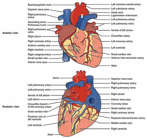
Figuring Out Cardiac Anatomy: Your Heart - dummies In fact, your heart is situated slightly to the left of center in your chest. Figure 1: Anterior view of the heart. A thick layer of muscle tissue and a protective membrane that folds into two layers, called the pericardium or pericardial membranes, surround the heart. The heart itself is a well-organized grouping of hollow spaces.
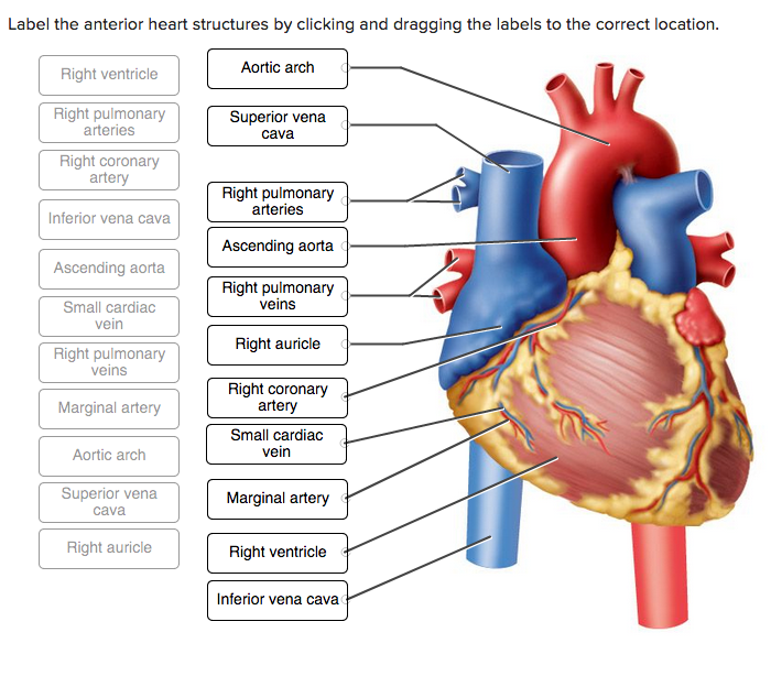
Solved Label the anterior heart structures by clicking and ...
Heart Labeling Quiz: How Much You Know About Heart Labeling? Here is a Heart labeling quiz for you. The human heart is a vital organ for every human. The more healthy your heart is, the longer the chances you have of surviving, so you better take care of it. Take the following quiz to know how much you know about your heart. Questions and Answers. 1.

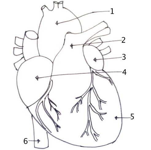
Q4 Given alongside is a diagram of the human heart showing ...
en.wikipedia.org › wiki › SGLT2_inhibitorSGLT2 inhibitor - Wikipedia The structure-activity relationship (SAR) of gliflozins is not fully understood. The most common gliflozins are dapagliflozin, empagliflozin and canagliflozin. The differences in the structures is relatively small. The general structure includes a glucose sugar with an aromatic group in the β-position at the anomeric carbon.

Structure of the heart. | Download Scientific Diagram
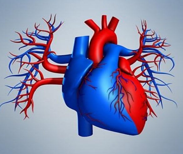
circulatory system diagram without labels system circulatory labels google. 174 Best Circulatory System Images On Pinterest | Human Anatomy, The . circulatory system human diagram nursing body anatomy medical circulation arteries cardiac heart systems vessels cardiovascular science simple enlarge major blood. Circulatory System ~ Nursing nursing-skills.blogspot.com
Easy trick to draw Human Heart
diagram of heart without labels 13 Best Images of Hip Anatomy Of The Worksheet - Sunflower Anatomy. 11 Pics about 13 Best Images of Hip Anatomy Of The Worksheet - Sunflower Anatomy : u414adad: heart diagram without labels, Congestive Heart Failure: The Essence of Heart Failure Course | CEUfast and also 13 Best Images of Hip Anatomy Of The Worksheet - Sunflower Anatomy.

Label every structure on the figure of the heart: image ...
professional.heart.org › en › partnersFellow of the American Heart Association (FAHA) International applicants will be required to provide the same online application data as domestic applicants. Letter of Recommendation. If the candidate does not have access to a FAHA who knows their work, their letter of recommendation may be authored by the Chair or Academic Chair of their institution or by an international scientific leader.
The Heart | Circulatory Anatomy
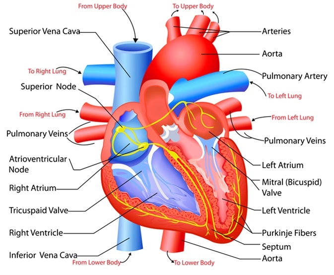
Heart Diagram with Labels and Detailed Explanation - BYJUS Diagram of Heart. The human heart is the most crucial organ of the human body. It pumps blood from the heart to different parts of the body and back to the heart. The most common heart attack symptoms or warning signs are chest pain, breathlessness, nausea, sweating etc. The diagram of heart is beneficial for Class 10 and 12 and is frequently ...
Heart dissection - BIOLOGY4ISC
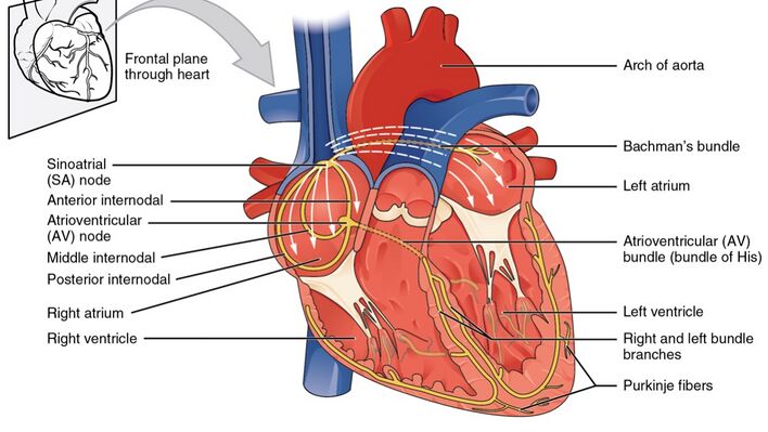
CH. 20 Assessment Flashcards | Quizlet Place the numbers in the locations corresponding to the correct sequence of blood flow through the heart, beginning with the superior vena cava. Correctly label the following external anatomy of the anterior heart. Place the components of the electrical conducting system in order from the initiation of an action potential until the end.

Heart Structure | BioNinja
Anatomy of a Human Heart - U of M Health Located between the lungs in the middle of the chest, the heart pumps blood through the network of arteries and veins known as the cardiovascular system. It pushes blood to the body's organs, tissues and cells. Blood delivers oxygen and nutrients to every cell and removes the carbon dioxide and other waste products made by those cells.

Simple Heart Diagram label | School | Clipart library - Clip ...
Diagram Of Fish With Label / External Morphology Of Rohu Fish With ... Plain diagram of the heart with labels to add and a cloze exercise on the pathway of blood through the heart. Structure of a typical fish (with diagram). A quality educational site offering 5000+ free printable theme units, word puzzles, writing forms, book report forms,math, ideas, lessons and much more.

Heart Anatomy: Labeled Diagram, Structures, Blood Flow ...
Structure and Function of the Heart - News-Medical.net Structure of the heart, The heart wall is composed of three layers, including the outer epicardium (thin layer), middle myocardium (thick layer), and innermost endocardium (thin layer). The...
The Human Heart

Diagram Of The Heart No Labels, HD Png Download , Transparent ...
Free Heart Diagram Unlabeled, Download Free Heart Diagram ...
13+ Heart Diagram Templates – Sample, Example, Format ...
Anatomy of the Human Heart - Physiopedia

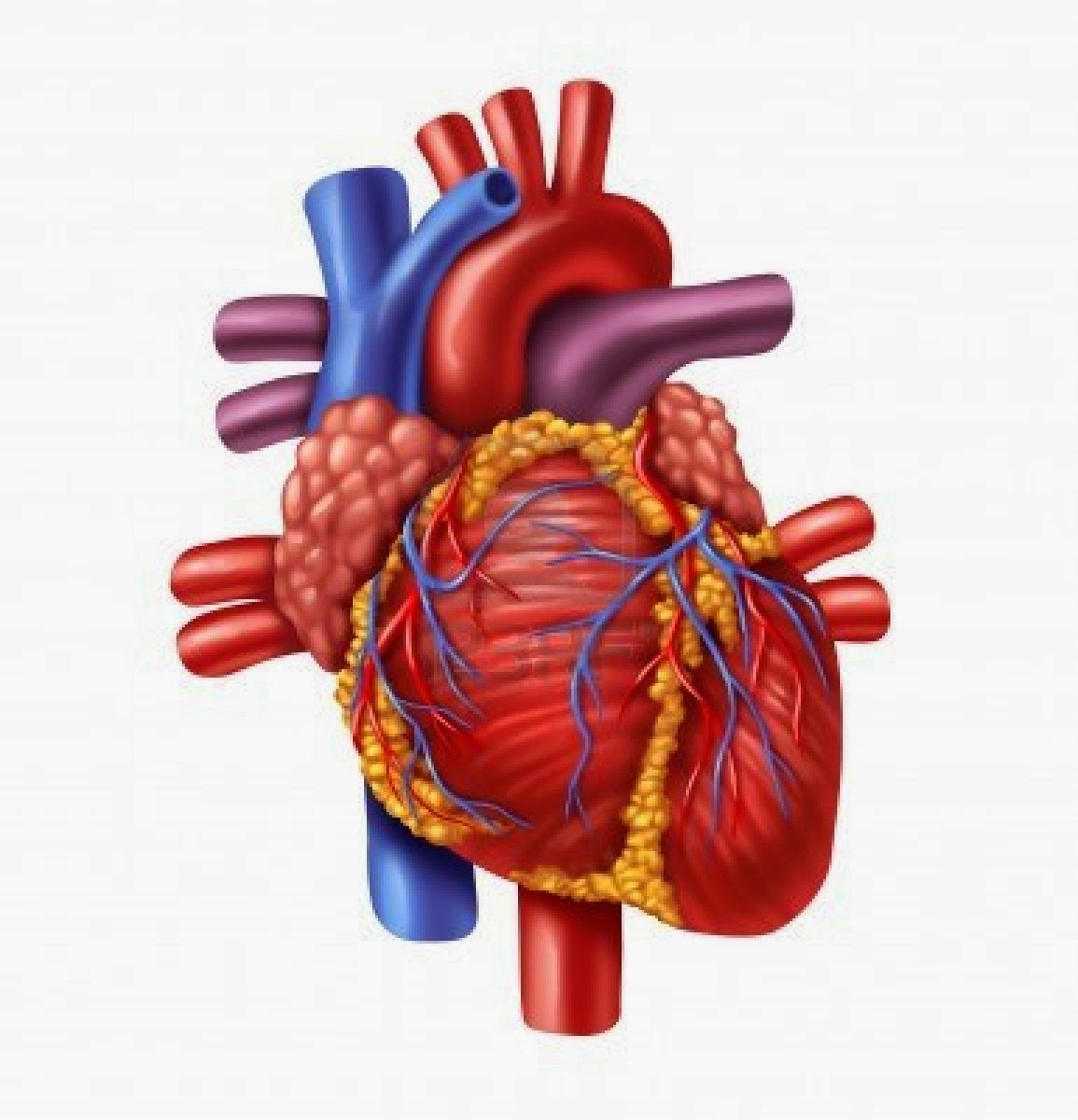
Human Heart Anatomy Stock Illustrations – 32,278 Human Heart ...

40.9: Mammalian Heart and Blood Vessels - Structures of the ...
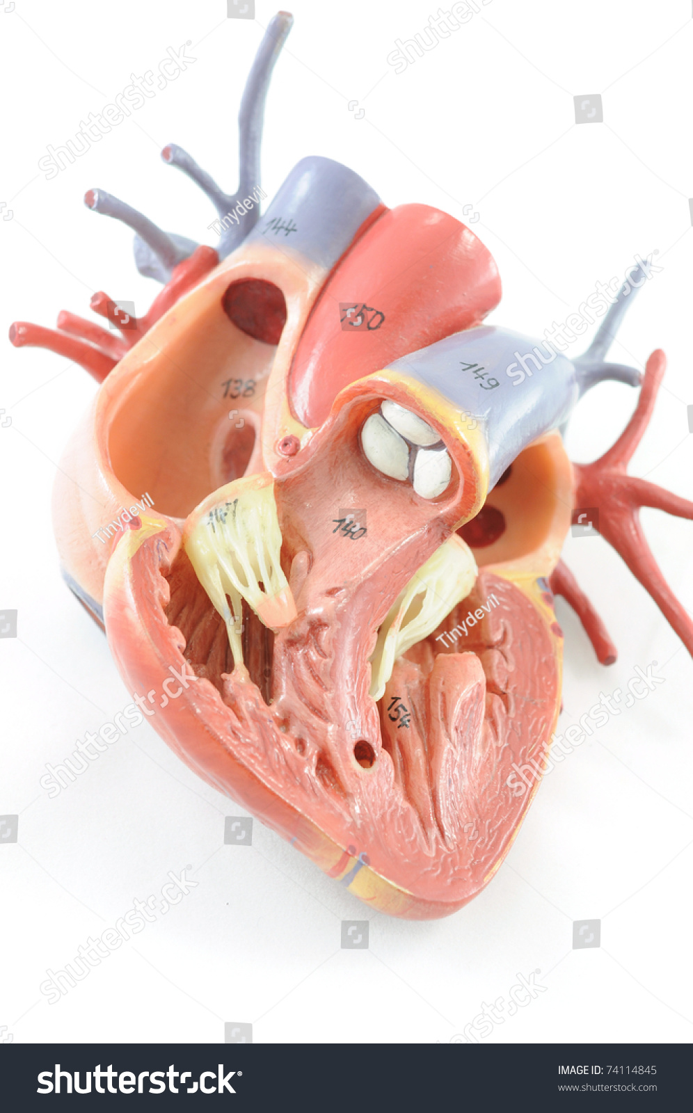
Close Human Heart Anatomy Stock Photo 74114845 | Shutterstock

How to draw internal structure of Human heart - Easy version ...

Sketch Of Human Heart Anatomy With Hand Written Labels Stock ...
Structure and Function of the Heart
Free Heart Diagram Unlabeled, Download Free Heart Diagram ...
Anatomy of the Human Heart - Physiopedia
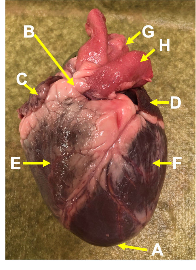
Heart Anatomy: Heart Dissection
Learn the Anatomy of the Heart

Draw labelled diagram of internal structure of human heart ...

Heart Structure | BioNinja
Heart: Anatomy | Concise Medical Knowledge
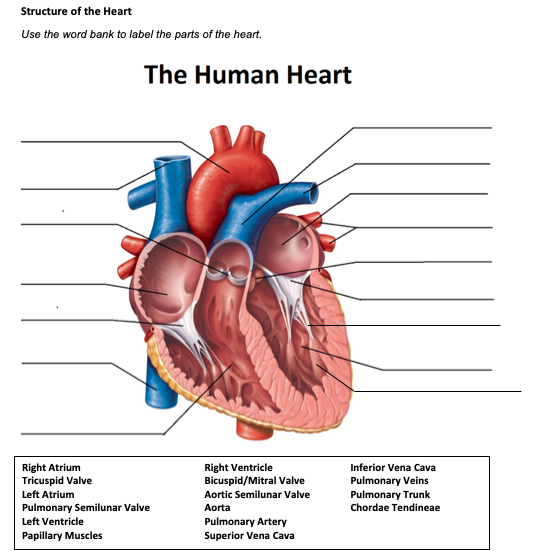
Solved Structure of the Heart Use the word bank to label the ...
Heart Structure - Ms. Campbell
Free Heart Diagram Unlabeled, Download Free Heart Diagram ...
Heart Anatomy: Labeled Diagram, Structures, Blood Flow ...
Heart Structure Without Label, HD Png Download - kindpng

Anatomy of a Human Heart

Heart Anatomy | Anatomy and Physiology II
Notes: Heart and Circulatory System

Label this: posterior surface heart structures Diagram | Quizlet
The Structure of the Heart Learning Objectives: Label the ...
Anatomy of the Human Heart - Physiopedia
13+ Heart Diagram Templates – Sample, Example, Format ...
Post a Comment for "42 structure of the heart without labels"