39 onion cells under microscope with labels
Observing Onion Cells Under The Microscope Afterwards, carefully mount the prepared and stained onion cell slide onto the microscope stage. Make sure that the cover slip is perfectly aligned with the microscope slide, and that any excess stain has been wiped off. Secure the slide on the stage using the stage clips. Cell Under Microscope Onion Labeled observe the onion under the low and high power of your microscope label the cell wall, cytoplasm, and the pigmented organelle structures (but you must label it with their real name using the evidence 1 look at each slide of specialised cells 04" (75mmx25mmx1mm) manufactured under iso 9001 quality control standard the diagram below shows onion …
Microscope Cell Lab: Cheek, Onion, Zebrina - SchoolWorkHelper The onion epidermis cell is the only cell that has a cell wall. In addition, it is the only cell that has a chloroplast, where photosynthesis can happen. The cheek epithelium cell is the only one that has centrioles, the barrel-shaped organelle that is responsible for helping organize chromosomes during cell division.

Onion cells under microscope with labels
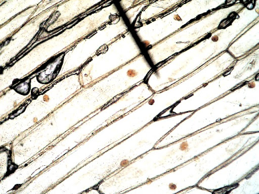
DOC The Onion Cell Lab - chsd.us Onion tissue provides excellent cells to study under the microscope. The main cell structures are easy to see when viewed with the microscope at medium power. For example, you will observe a large circular . nucleus. in each cell, which contains the genetic material for the cell. In each nucleus, are round bodies called . nucleoli PDF Onion Cells - Investigation - Exploring Nature 5. Observe the onion tissue under the microscope at 4x, 10x and 40x with lots of light (open diaphragm). Then slowly close the diaphragm while observing the image to find the best light for seeing cellular details. 6. Draw a section of onion skin cells at 10x magnification. Then switch to 40x and draw one cell and label it. Questions: 1. Onion Epidermis - kuensting.org Onion epidermal cells, iodine stain, 400X. The nucleus of an onion epidermal cell, 1000X magnification. ...
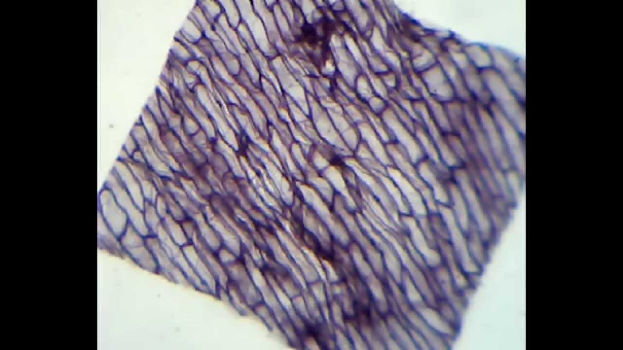
Onion cells under microscope with labels. Onion Cell Lab Report.docx - Onion Cell Lab Report By - Course Hero Onion Cell Lab Report By : Nawaf Almalki Introduction: Many things that are viewed using a microscope, particularly cells, can appear quite transparent under the microscope. The internal parts of the cells, the organelles, are so transparent that they are often difficult to see. Biologists have developed a number of stains that help them see the cells and their organelles by adding color to ... Under the Micrsocope: Onion Cell (100x - 400x) - YouTube In this "experiment" we will see onion cells under the microscope.For the experiment you will only need onion, dropper and the microscope (container and tool... Onion Skin Cells - Investigation - Exploring Nature 5. Observe the onion tissue under the microscope at 4x, 10x and 40x with lots of light (open diaphragm). Then slowly close the diaphragm while observing the image to find the best light for seeing cellular details. 6. Draw a section of onion skin cells at 10x magnification. Then switch to 40x and draw one cell and label it. Onion Plant Cell Under Microscope Labeled - Ismael Dauila Explore diffusion/osmosis by looking at onion cells under the microscope. It is used for treating a parasite disease called ich (ichthyophthirius multifiliis; Label the cell wall and chloroplasts. Students will observe plant cells using a light microscope.
Looking at the Structure of Cells in the Microscope Both types of light microscopy are widely used to visualize living cells. Figure 9-7 Two ways to obtain contrast in light microscopy. (A) The stained portions of the cell reduce the amplitude of light waves of particular wavelengths passing through them. A colored image of the cell is thereby obtained that is visible in the ordinary way. (more...) What Does a Worm Look Like Under a Microscope? Place the leaves in a glass container with water for 30 minutes. After 30 minutes, you'll notice eelworms (in a group) crawling around the bottom of the container. Take off the water carefully, avoiding the eelworm mass. You can keep the eelworms in the jar or gently pour them into a Petri dish with a bit of water. Onion Skin Cells Labeled - the wonderful microworld onion skin cells ... Onion Skin Cells Labeled. Here are a number of highest rated Onion Skin Cells Labeled pictures on internet. We identified it from obedient source. Its submitted by executive in the best field. We... Onion Cell Diagram Labeled Pdf .pdf - thesource2.metro Set your multimeter to measure current in the 20 mA range (the dial setting labeled "20m" on the right). Plug the multimeter's black probe into the port labeled COM. Plug the multimeter's red probe into the port labeled VΩmA. Use a red alligator clip lead to connect the multimeter's red probe to the positive (+) terminal of the 9 V battery.
1,922 Onion cell Images, Stock Photos & Vectors - Shutterstock 1,922 onion cell stock photos, vectors, and illustrations are available royalty-free. See onion cell stock video clips Image type Orientation People Artists Sort by Popular Food and Drink Science Biology garden onion microscope cell photomicrography mitotic cell cycle epidermi optical microscope Next of 20 Onion Cells Under a Microscope (100x-2500x) - YouTube In this video you will see onion cells under a microscope (100x-2500x) as is, without any coloring. To observe the onion cells the thin membrane is used. It... Onion Cells Under a Microscope - Requirements/Preparation/Observation Add a drop of iodine solution on the onion membrane (or methylene blue) Gently lay a microscopic cover slip on the membrane and press it down gently using a needle to remove air bubbles. Touch a blotting paper on one side of the slide to drain excess iodine/water solution, Place the slide on the microscope stage under low power to observe. DOC Plant and Animal Cells Microscope Lab - Hillsboro City Schools Make a drawing of one onion cell, labeling all of its parts as you observe them. (At minimum you should observe the nucleus, cell wall, and cytoplasm.) Cheek cells 1. To view cheek cells, gently scrape the inside lining of your cheek with a toothpick. DO NOT GOUGE THE INSIDE OF YOUR CHEEK! (We will observe blood cells in a future lab!!) 2.
Rens blog : March 2014
Under Cell Cheek Microscope Human The cheek cells were scraped from the mouth and were examined as follows: compare and contrastmale and female cheek cells , emerging from the surface of cells (green) cultured in the lab diagram onion cells and label the cell wall, cell membrane, cytoplasm, nucleus, and chloroplast cell walls and chloroplasts 3 cell walls and chloroplasts 3.
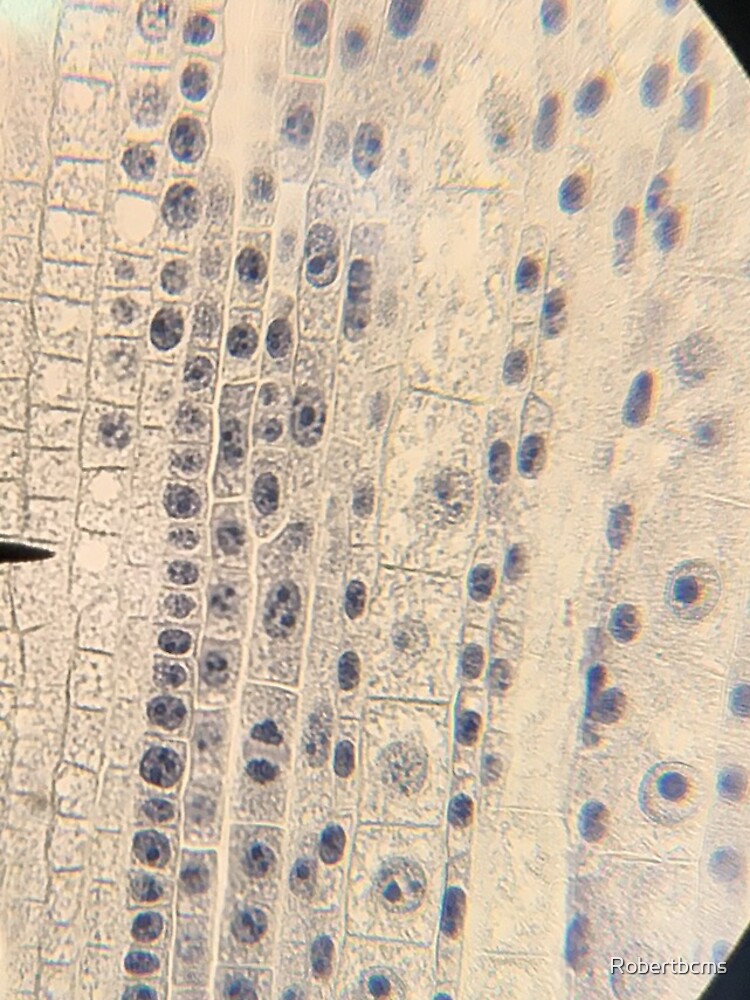
"Biology- onion root tip cells under microscope" iPhone Case & Cover by Robertbcms | Redbubble
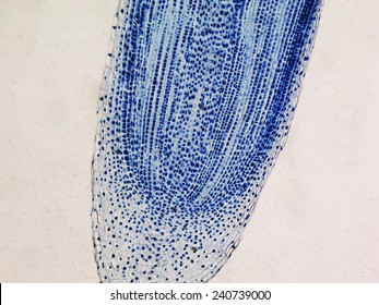
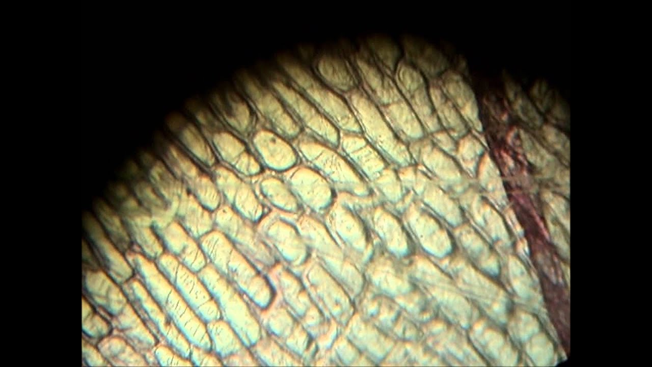
Onion Root Mitosis - Microscopy-UK Onions have larger chromosomes than most plants and stain dark. The chromosomes are easily observed through a compound light microscope. The cells pictured below are located in the apical meristem of the onion root. The apical meristem is an area of a plant where cell division takes place at a rapid rate. Phases of plant cells division:
Onion cells under the microscope: 40X - 100X - 400X - YouTube
Onion Root Tip Mitosis - Stages, Experiment and Results The cellulose produced by the two new cells occupies the region between the middle lamella and cell membrane to form the primary cell wall for the two daughter cells. Microscope Experiments. Difference between Meiosis and Mitosis. Return to Onion Cells under the Microscope. Return from Onion Root Tip Mitosis to Microscopemaster home
Onion cells under a microscope 400x 1000x
Plant Cell Under Microscope Labeled 40X : Young Root 2 Of Broad Bean ... Microscopy and the interpretation of cell structures. (iv) describe how you applied the stain. Students will observe cheek cells under a microscope. Does anyone have a decent labelled diagram of a plant cell under an electron microscope? Onion cell) magnification (40x, 100x, or 400x) label all visible cell parts *use pencil or colored pencil.

Biology 1 - Onion cell microscopy - YouTube
Microscopy, size and magnification - Microscopy, size and ... - BBC Place cells on a microscope slide. Add a drop of water or iodine (a chemical stain). Lower a coverslip onto the onion cells using forceps or a mounted needle. This needs to be done gently to...

swifty science: onion cell lab
Labeled Cell Microscope Onion Under draw and label your observations: label the nucleus, cell wall, and cytoplasm of one onion cell add one drop of iodine to the onion peel sample and place a cover slip over the newly stained tissue you will begin by observing cork cells under the microscope although all the onion bulb cells and the leaves of the onion contain the same dna, explain …

Fanos' MCB Blog: Onion Skin
PDF Onion Cell Lab - somewaresinmaine.com Research Biology Onion Cell Lab page 1 of 3 Onion Cell Lab After you have completed the rest of this lab come back to this cover page DRAW & LABEL AN ONION CELL WITH ALL THE PARTS / ORGANELLES YOU OBSERVE UNDER 40X. Purpose: To observe and identify major plant cell structures and to relate the structure of the cell to its function. Materials: 1 ...

Rens blog : Science, cells
Onion Cell Labeled Under Microscope size of onion cell-1600/2=800 µm draw a cell of each of the required specimens and label correctly the parts seen under the microscope look at various specimens under the microscope and compare the parts of a cell found in plants and animals observing onion cells- this post gives step by step instructions on how to prepare onion cells for …

Scientific Videos: Onion Epidermis, Slides, Cells
What organelles are in an onion cell? - Biology Stack Exchange You cannot see most of these as they appear translucent as well as being too small to see under the light microscope. You need an electron microscope to view these. Note: chloroplasts are not present in an onion cell as it is not a photosynthesising cell. This is a typical onion cell slide with labels:
Onion Root Tip Cell Under Microscope Labeled - Micropedia
Onion Epidermis - kuensting.org Onion epidermal cells, iodine stain, 400X. The nucleus of an onion epidermal cell, 1000X magnification. ...

How to Observe Onion Cells under a Microscope | Things under a microscope, Science cells, High ...
PDF Onion Cells - Investigation - Exploring Nature 5. Observe the onion tissue under the microscope at 4x, 10x and 40x with lots of light (open diaphragm). Then slowly close the diaphragm while observing the image to find the best light for seeing cellular details. 6. Draw a section of onion skin cells at 10x magnification. Then switch to 40x and draw one cell and label it. Questions: 1.
Biology Pictures: Onion Cells under Microscope
DOC The Onion Cell Lab - chsd.us Onion tissue provides excellent cells to study under the microscope. The main cell structures are easy to see when viewed with the microscope at medium power. For example, you will observe a large circular . nucleus. in each cell, which contains the genetic material for the cell. In each nucleus, are round bodies called . nucleoli

Cell structure | Cells as the basic units of life | Siyavula

Red Onion Cell Under Microscope Labeled - Micropedia

HOMESCHOOL NINJAS: Cell-U-Licious Saturday Science!

Onion root cells under MICROSCOPE - YouTube
Onion Cells Under Microscope! REALLY COOL!!! - YouTube
Post a Comment for "39 onion cells under microscope with labels"